Cell lysis is the first step in cell fractionation, organelle isolation and protein extraction and purification. As such, cell lysis opens the door to a myriad of proteomics research methods. Many techniques have been developed and used to obtain the best possible yield and purity for different species of organisms, sample types (cells or tissue) and target molecule or subcellular structure.
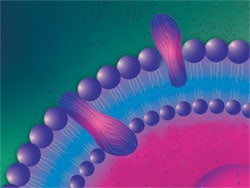
All cells have a plasma membrane, a protein-lipid bilayer that forms a barrier separating cell contents from the extracellular environment. Lipids comprising the plasma membrane are amphipathic, having hydrophilic and hydrophobic moieties that associate spontaneously to form a closed bimolecular sheet. Membrane proteins are embedded in the lipid bilayer, held in place by one or more domains spanning the hydrophobic core. In addition, peripheral proteins bind the inner or outer surface of the bilayer through interactions with integral membrane proteins or with polar lipid head groups. The nature of the lipid and protein content varies with cell type and species of organism.

Cell membrane structure. Illustration of a lipid bilayer comprising outer plasma membrane of a cell.
In animal cells, the plasma membrane is the only barrier separating cell contents from the environment, but in plants and bacteria the plasma membrane is also surrounded by a rigid cell wall. Bacterial cell walls are composed of peptidoglycan. Yeast cell walls are composed of two layers of ß-glucan, the inner layer being insoluble to alkaline conditions. Both of these are surrounded by an outer glycoprotein layer rich in the carbohydrate mannan. Plant cell walls consist of multiple layers of cellulose. These types of extracellular barriers confer shape and rigidity to the cells. Plant cell walls are particularly strong, making them very difficult to disrupt mechanically or chemically. Until recently, efficient lysis of yeast cells required mechanical disruption using glass beads, whereas bacterial cell walls are the easiest to break compared to these other cell types. The lack of an extracellular wall in animal cells makes them relatively easy to lyse.
There is no universal protocol for protein sample preparation. Sample preparation protocols must take into account several factors, such as the source of the specimen or sample type, chemical and structural heterogeneity of proteins, the cellular or subcellular location of the protein of interest, the required protein yield (which is dependent on the downstream applications), and the proposed downstream applications. For instance, bodily fluids such as urine or plasma are already more or less homogeneous protein solutions with low enzymatic activity, and only minor manipulation is required to obtain proteins from these samples. Tissue samples, however, require extensive manipulation to break up tissue architecture, control enzymatic activity, and solubilize proteins.
The quality or physical form of the isolated protein is also an important consideration when extracting proteins for certain downstream applications. For instance, applications such as functional enzyme-linked immunosorbent assay (ELISA) or crystallography require not only intact proteins but also proteins that are functionally active or retain their 3D structure.

Examples of protein sources for sample collection. Proteins can come from many sources, including the following: native sources such as mammalian cell cultures, tissues or bodily fluids; overexpression in a model system such as bacteria, yeast, insect or mammalian cells; monoclonal antibodies from hybridoma cells; or plant cells used in agricultural biotechnology.